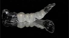
Multi-unit restorations
(Mandible /
Posterior)
複雑な部分欠損症例の治療
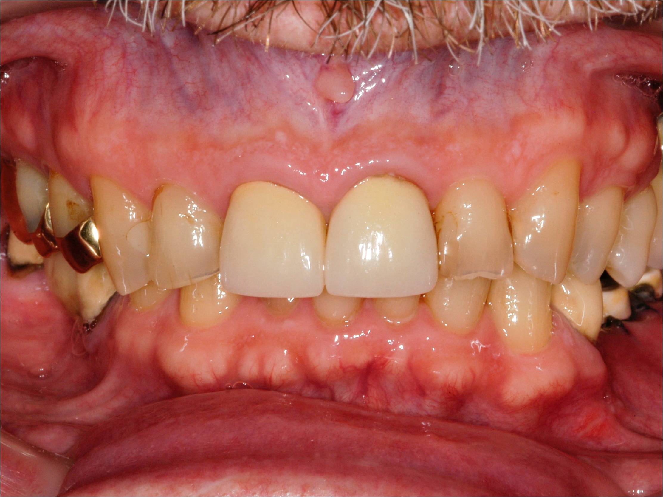
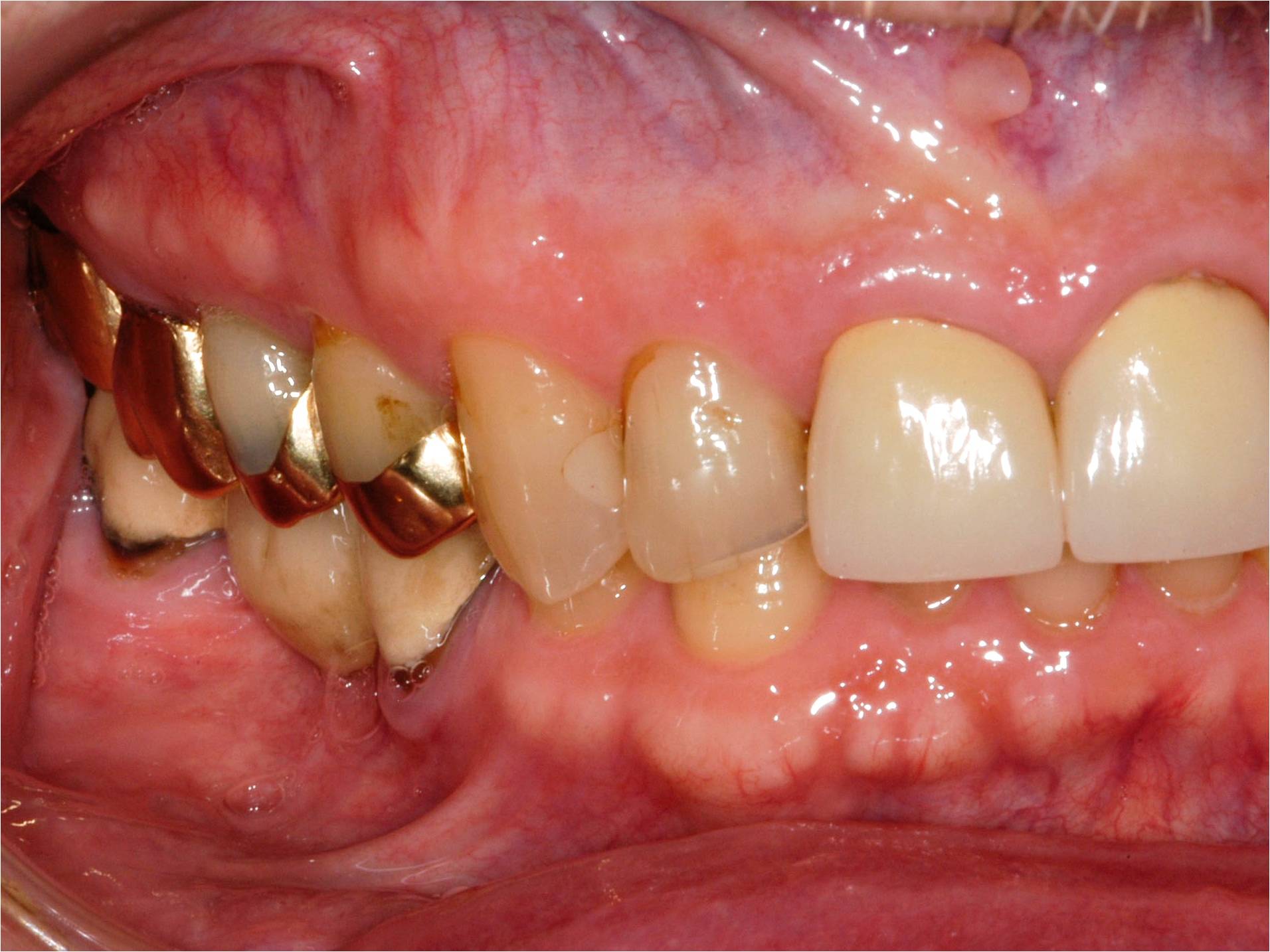
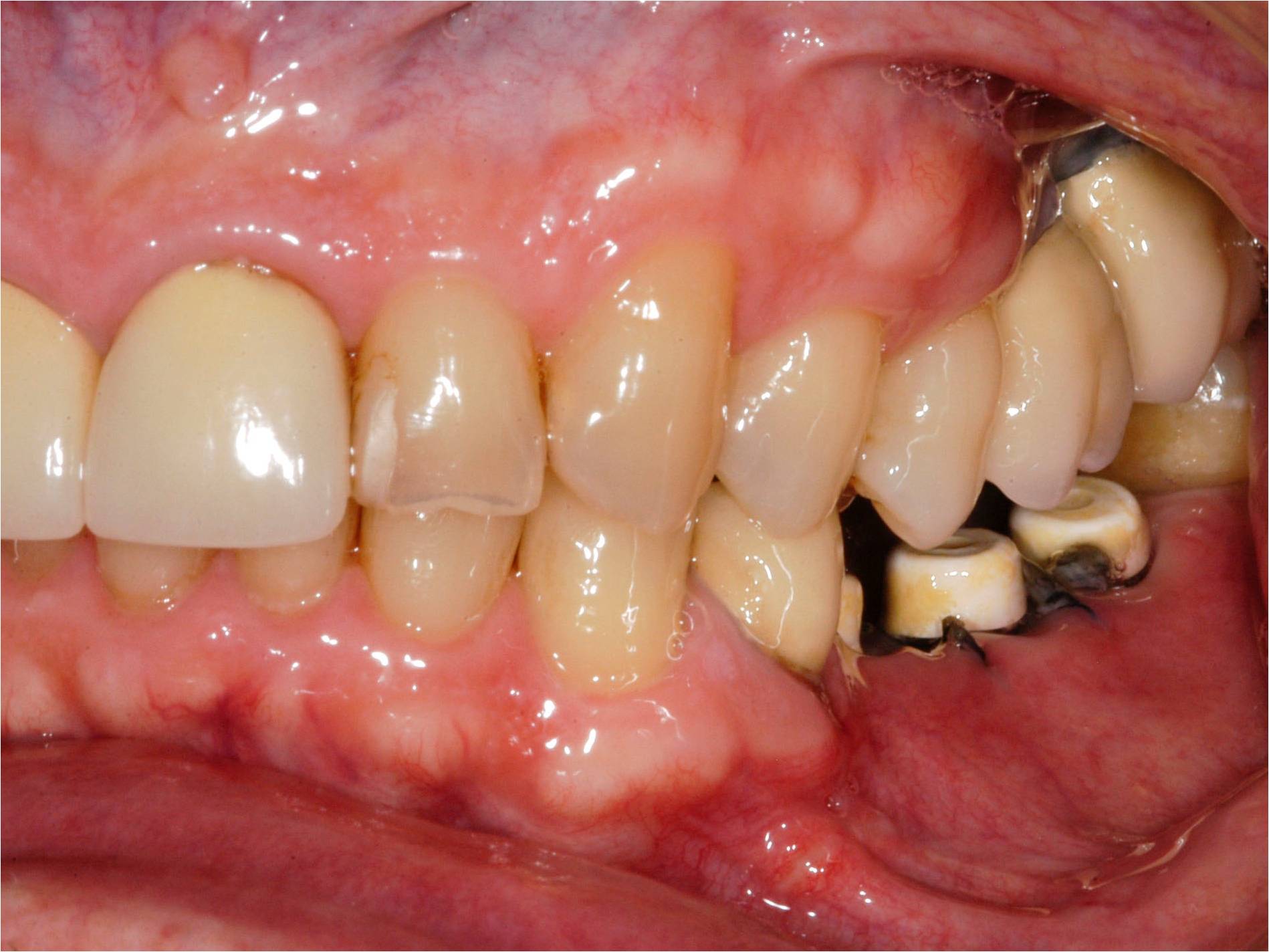
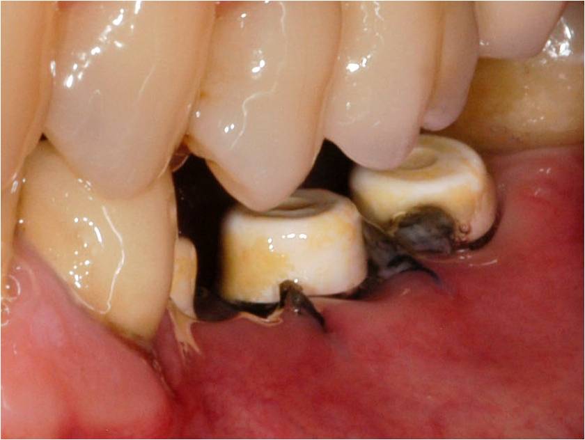
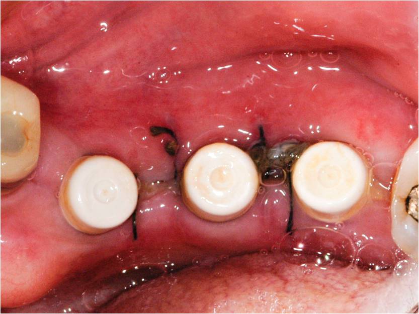
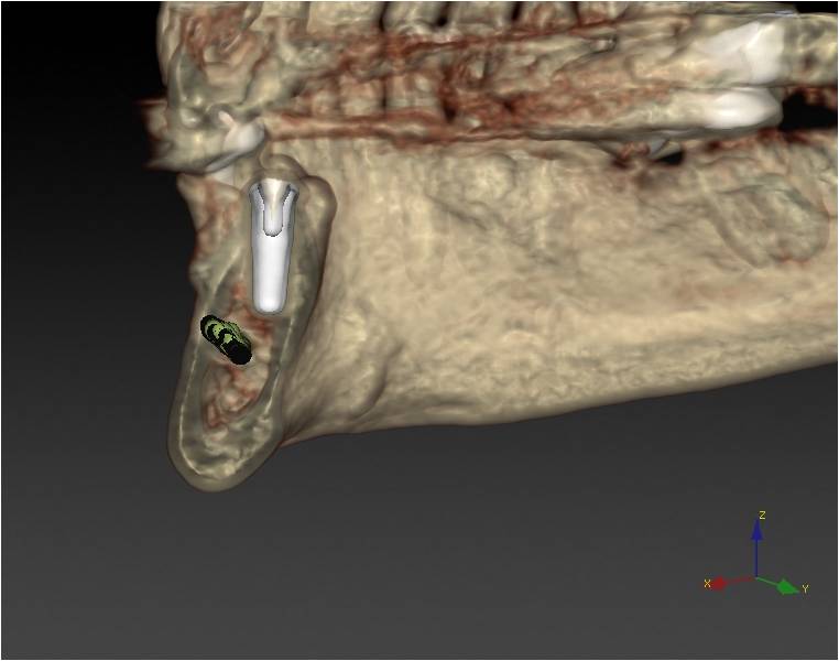
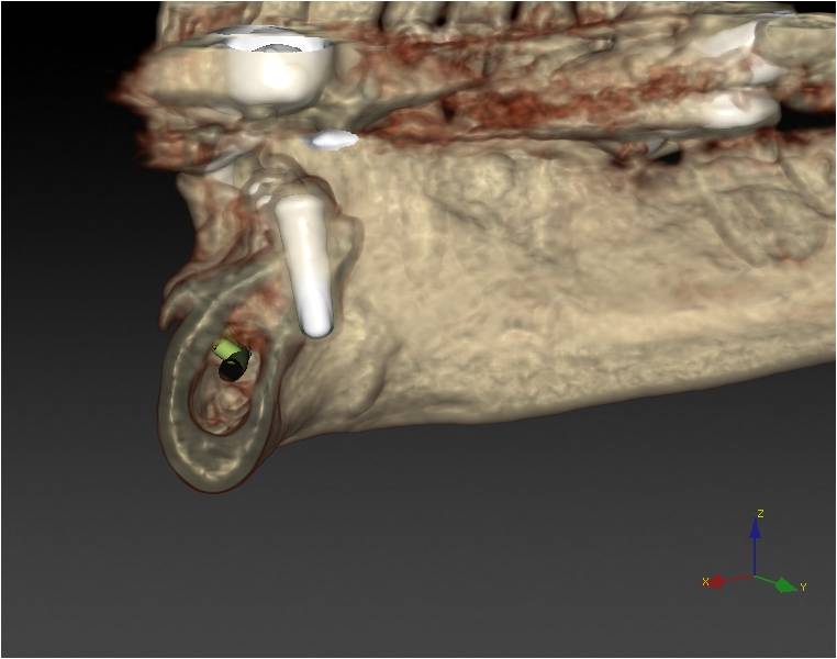
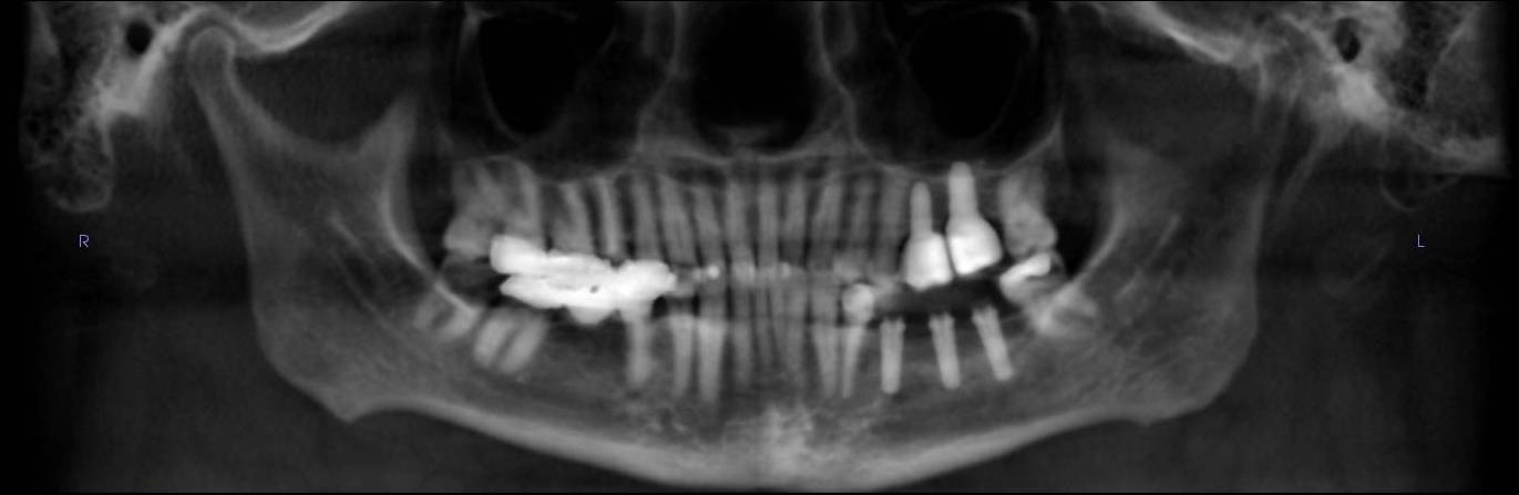
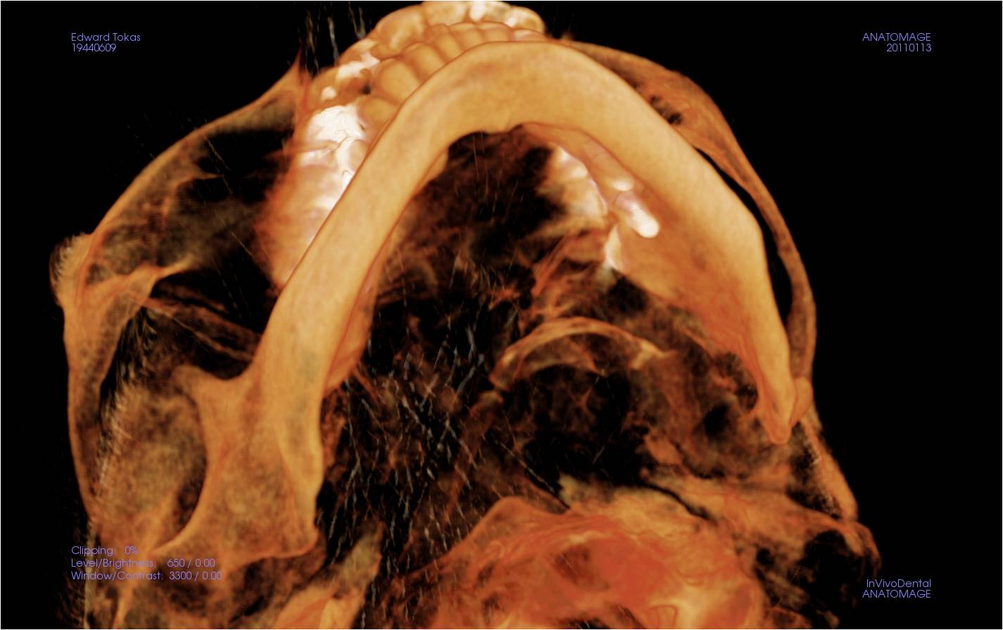
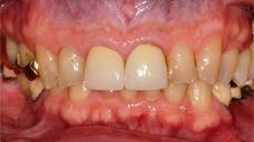
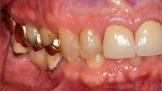
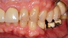
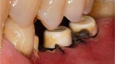
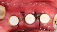
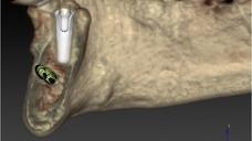
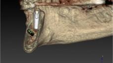
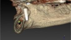
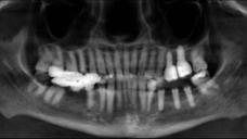
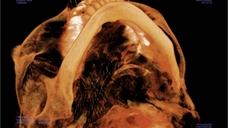
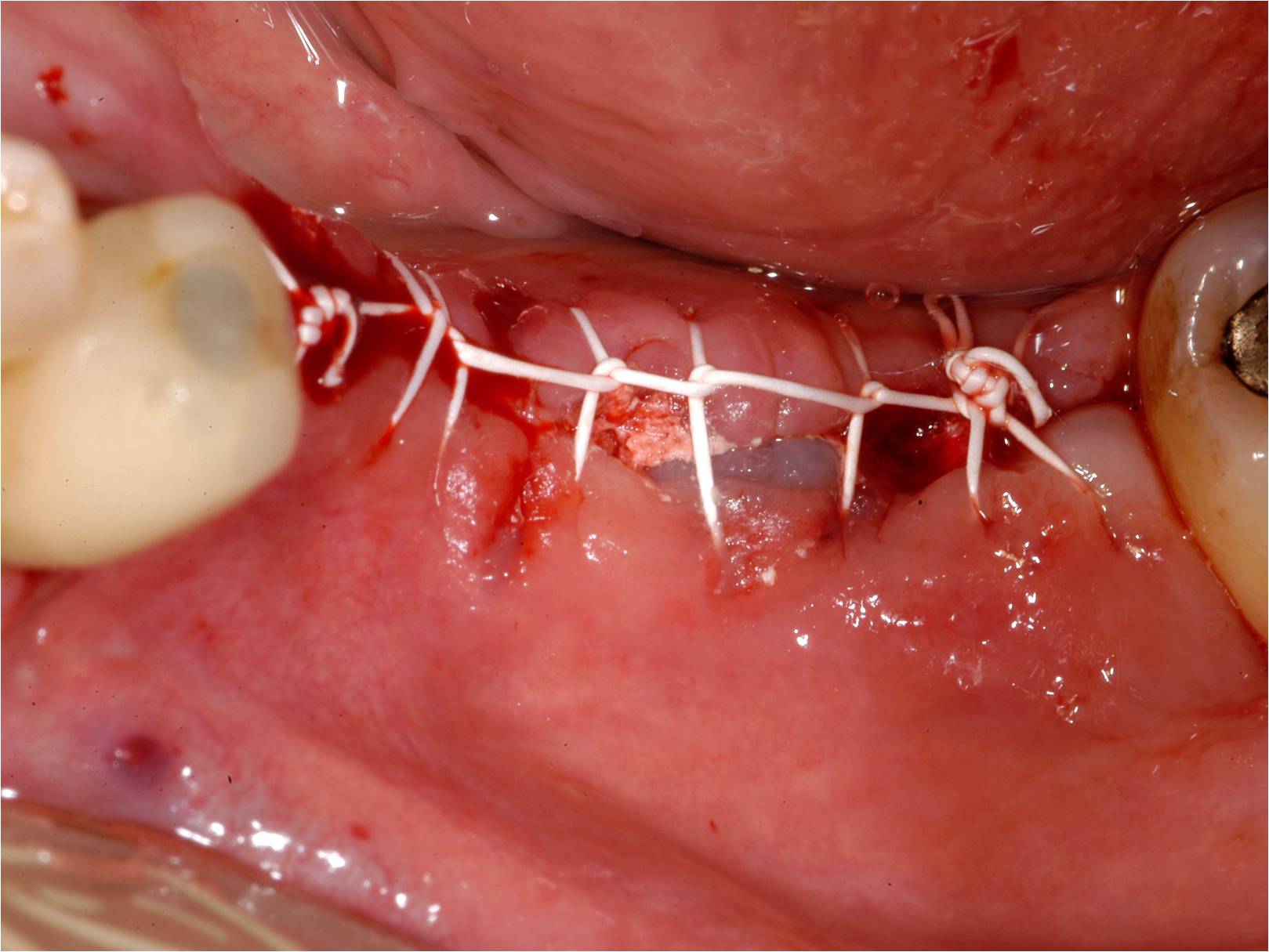
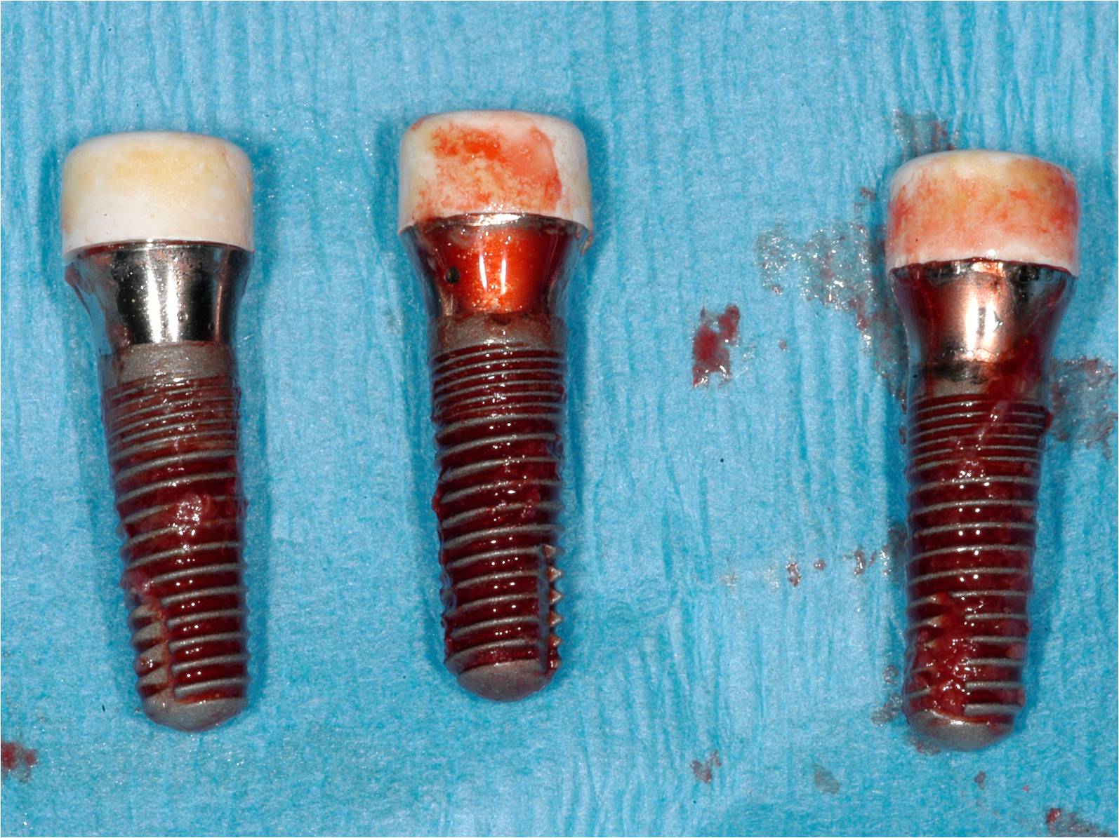
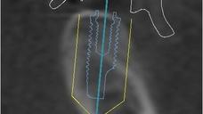
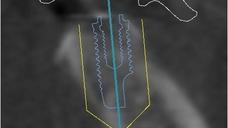
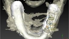
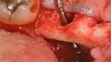
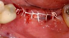
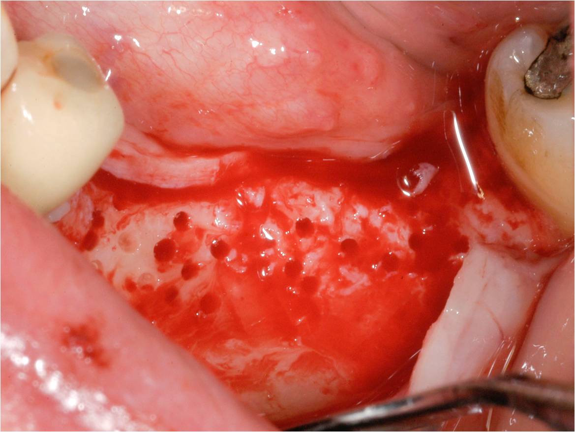
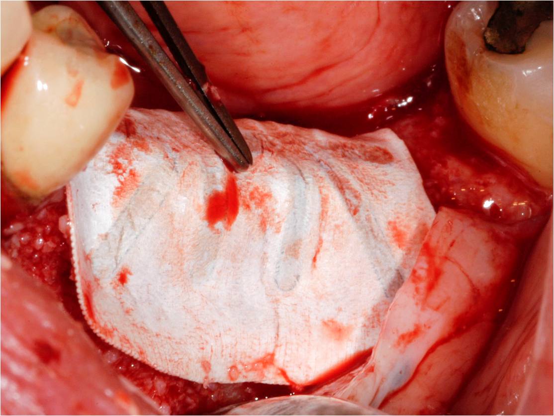
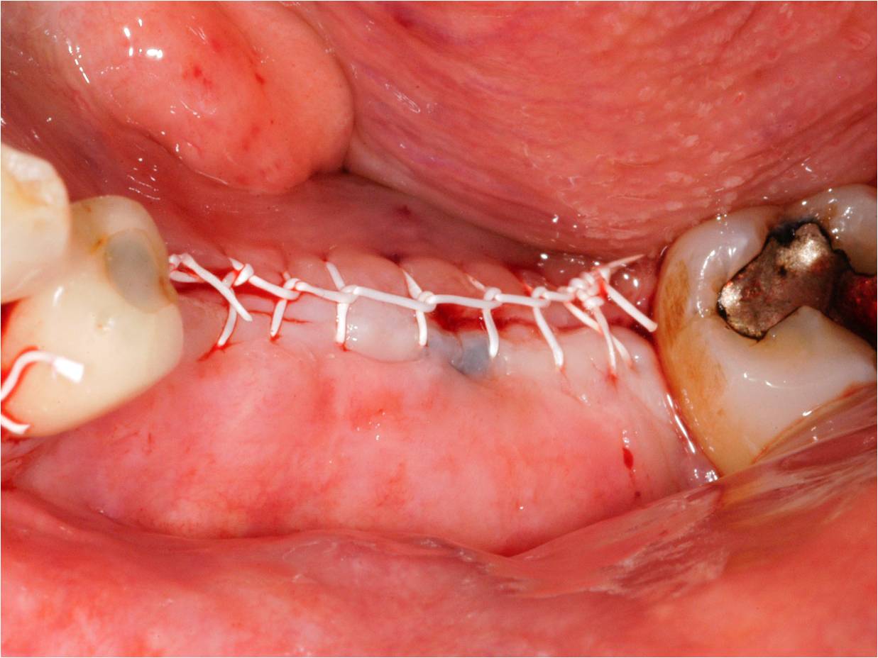
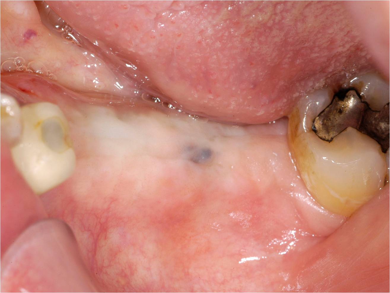
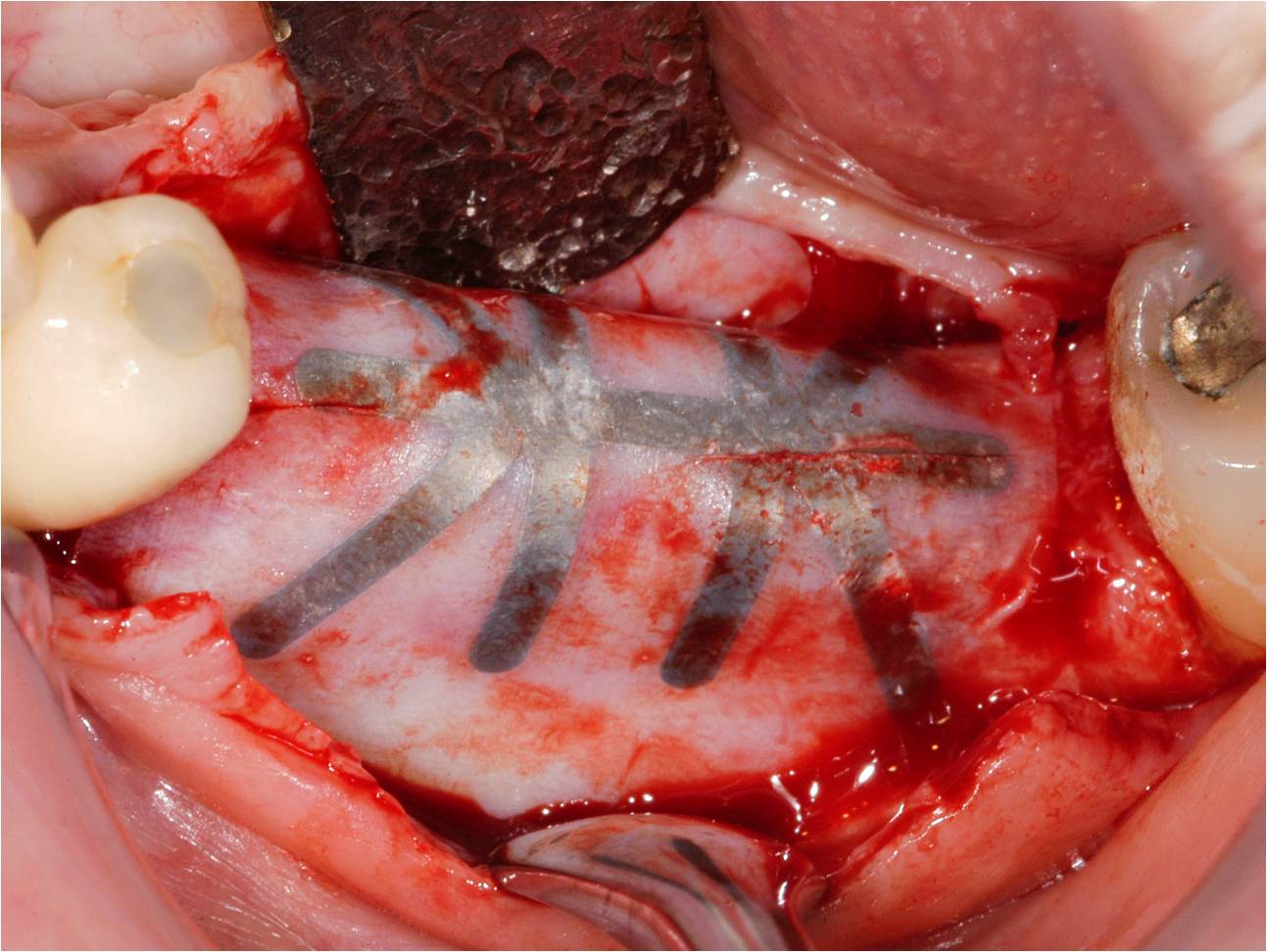
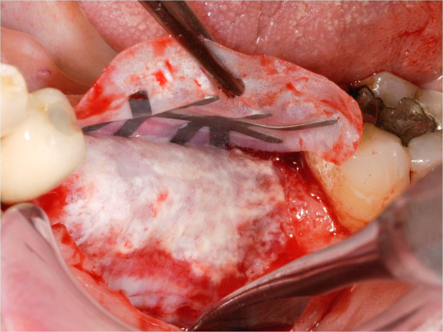
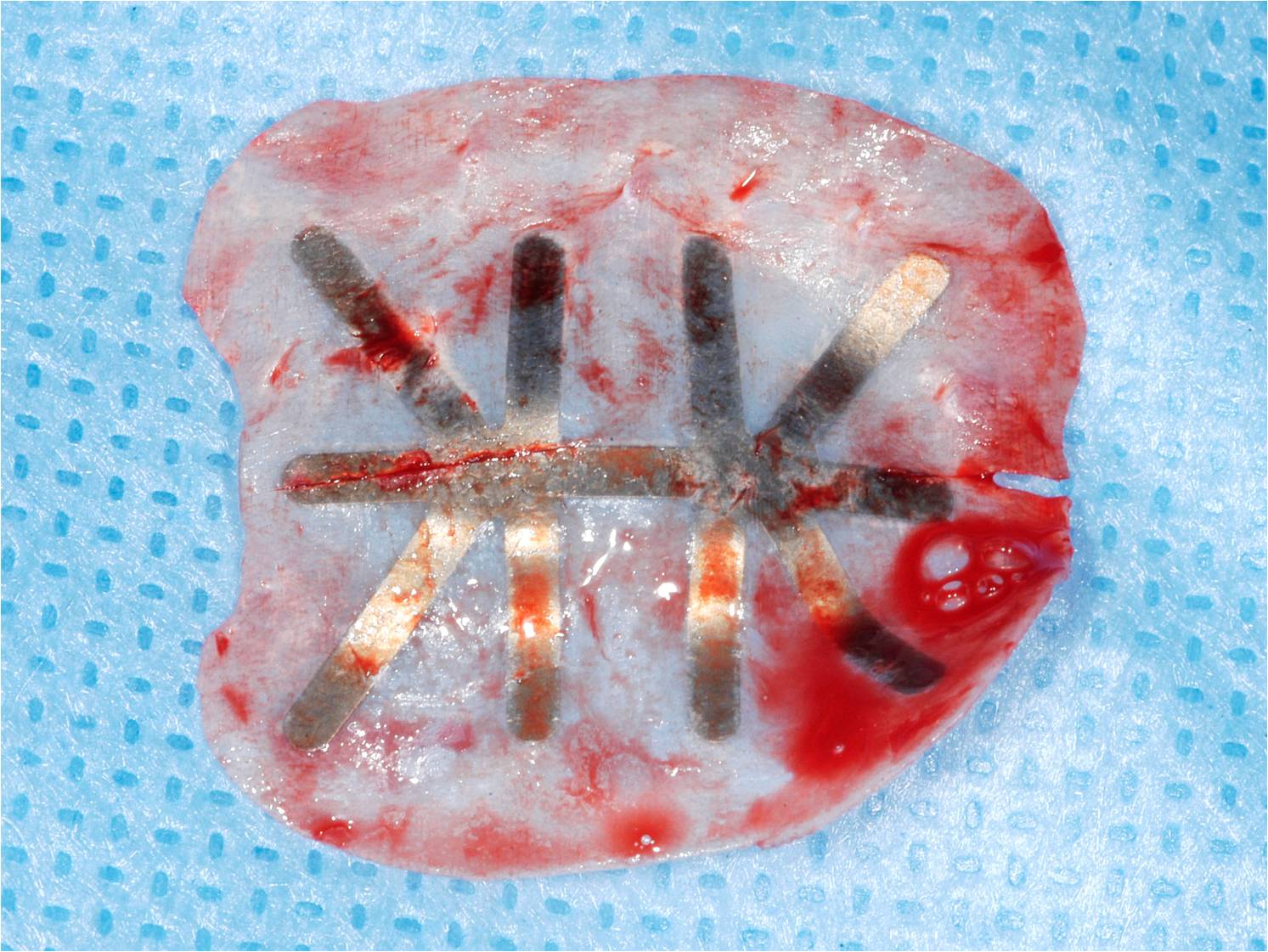
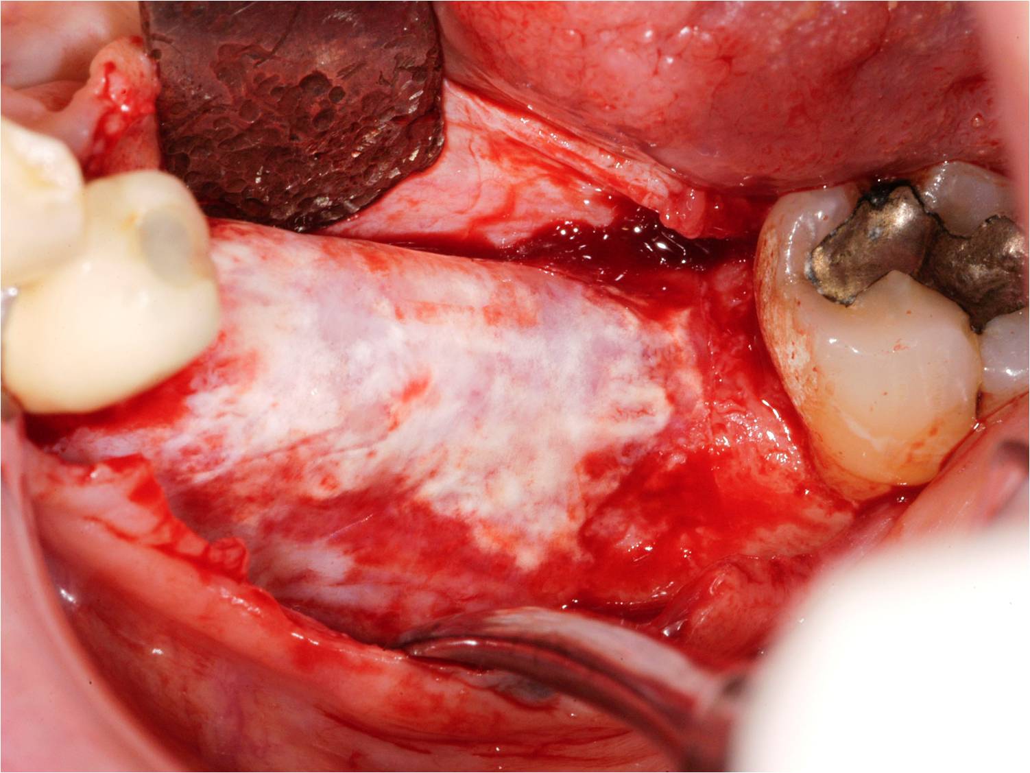
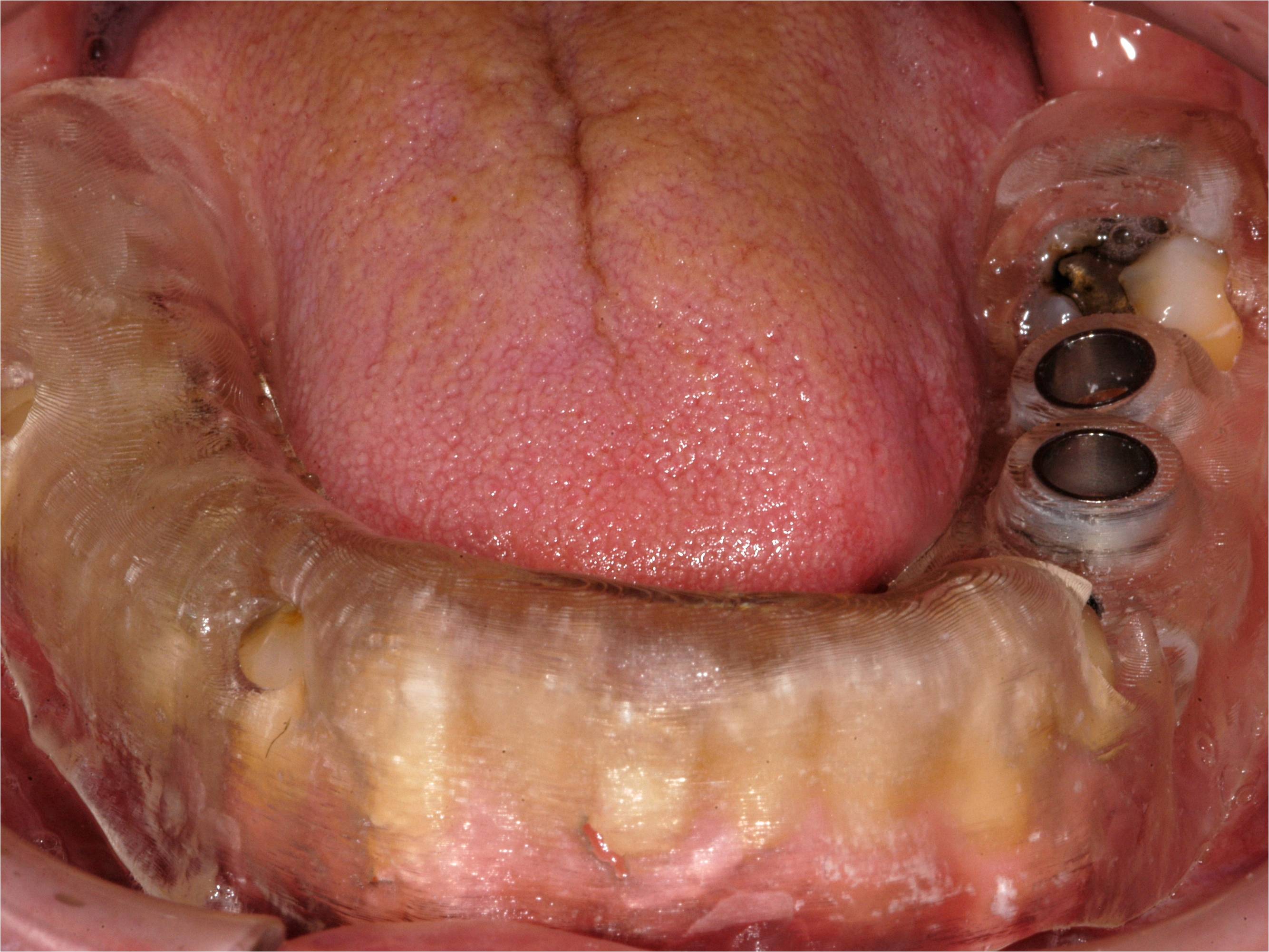
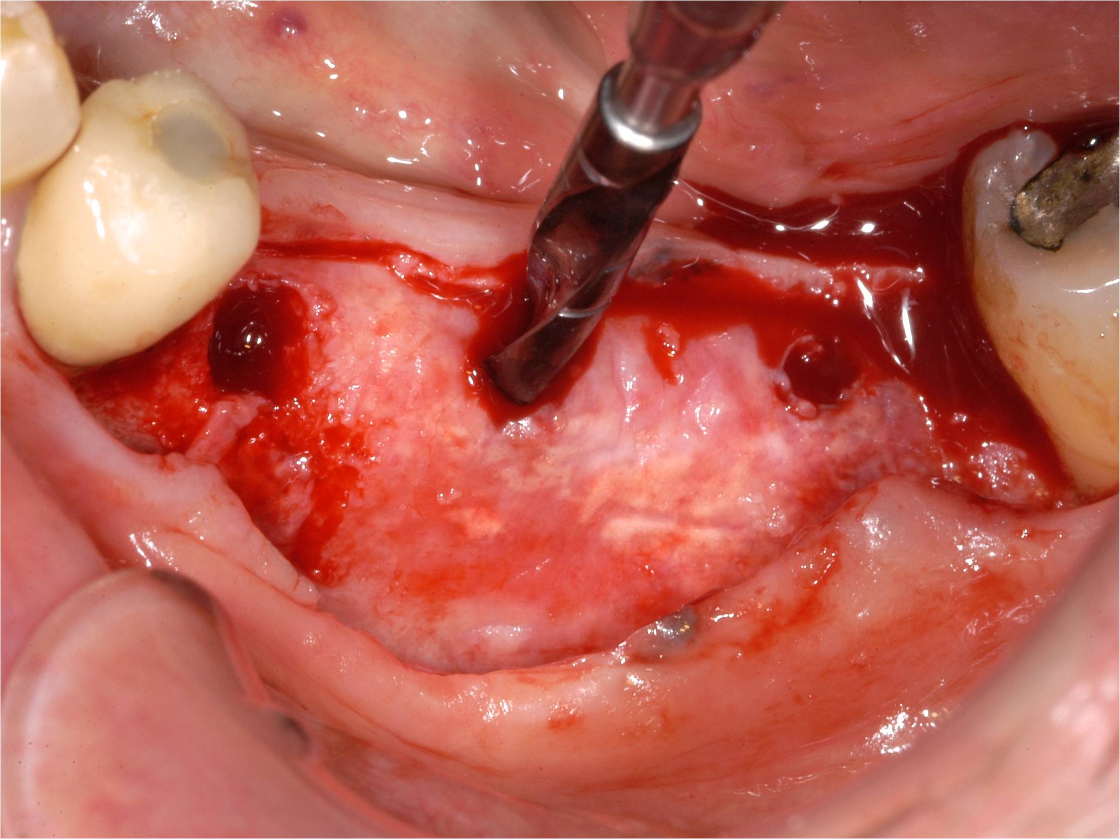
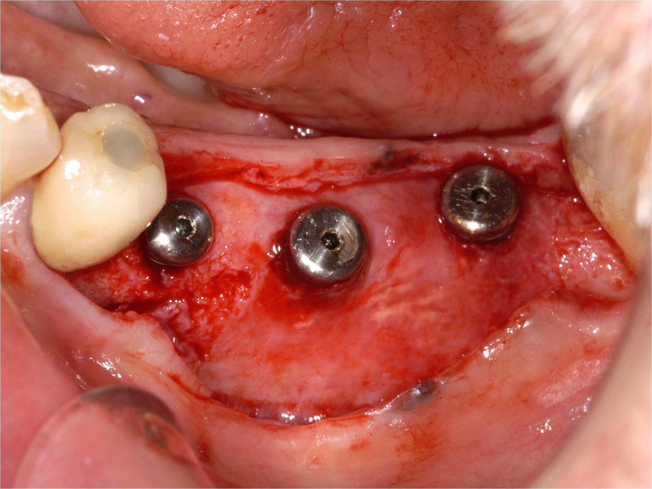
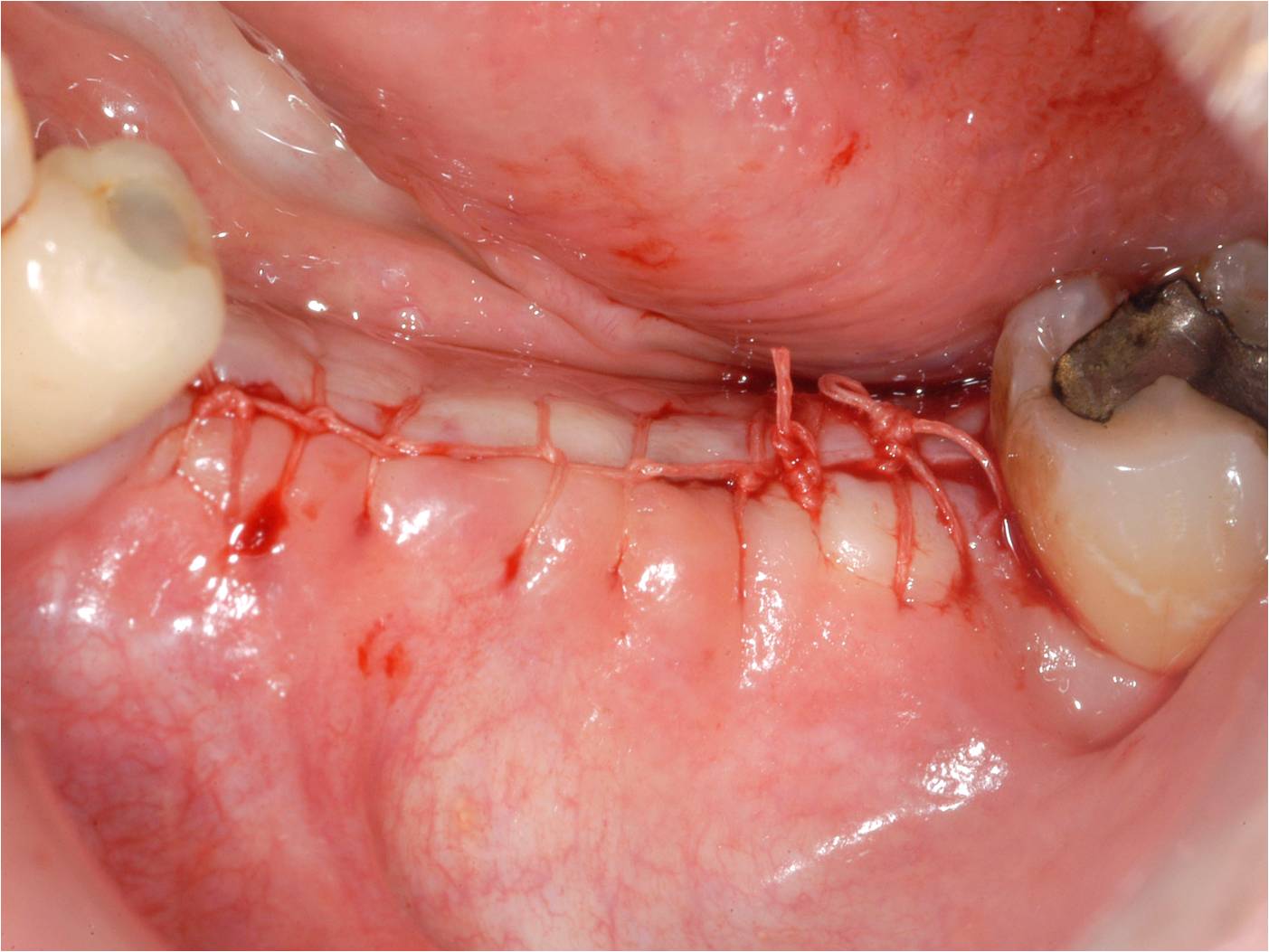
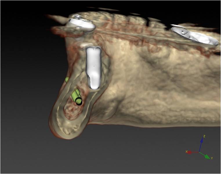
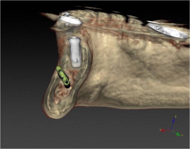
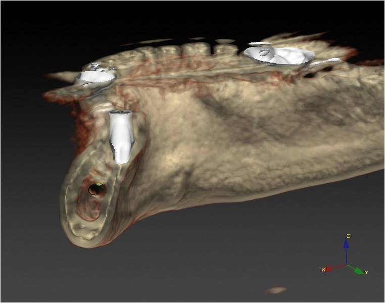
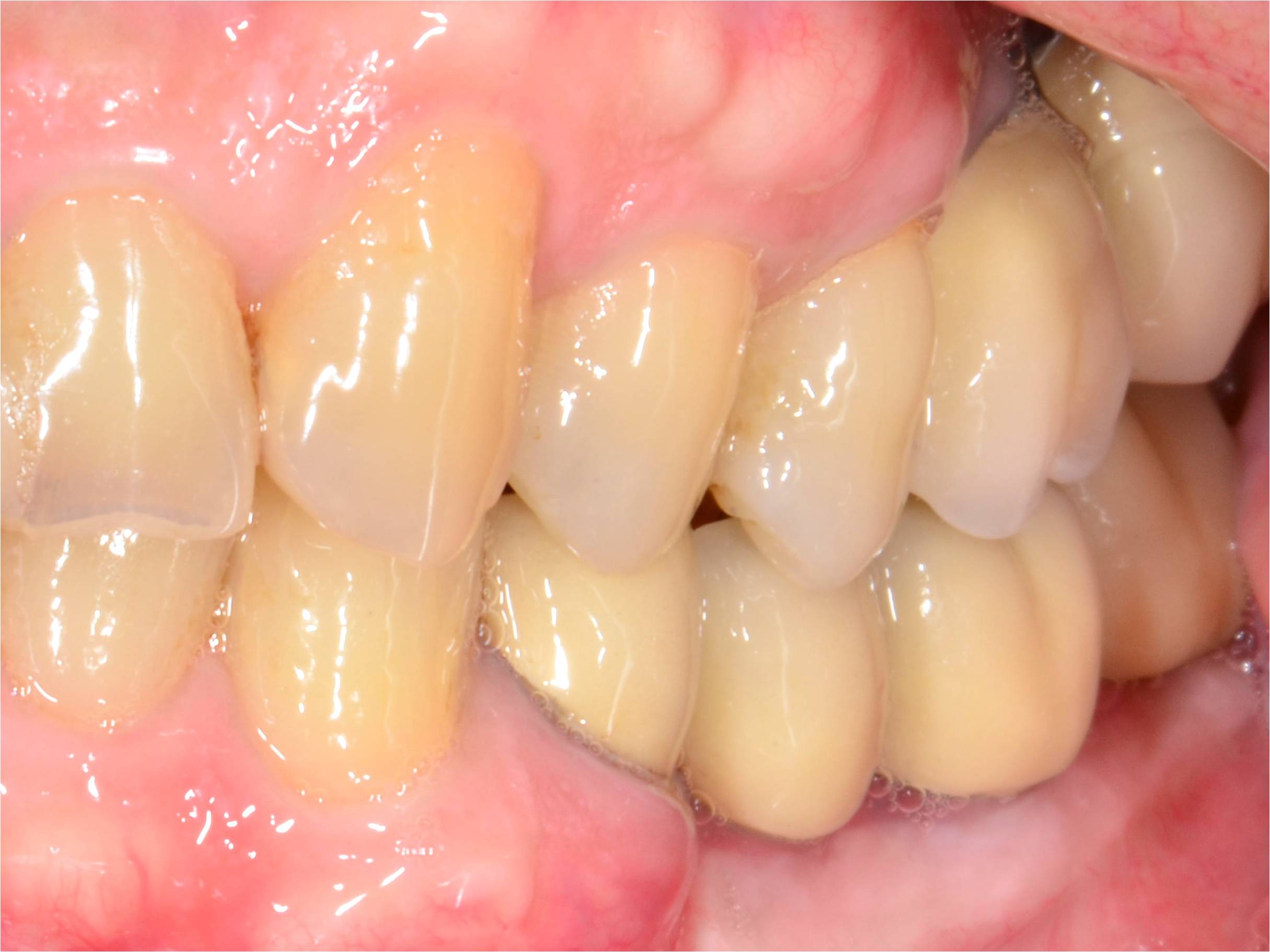
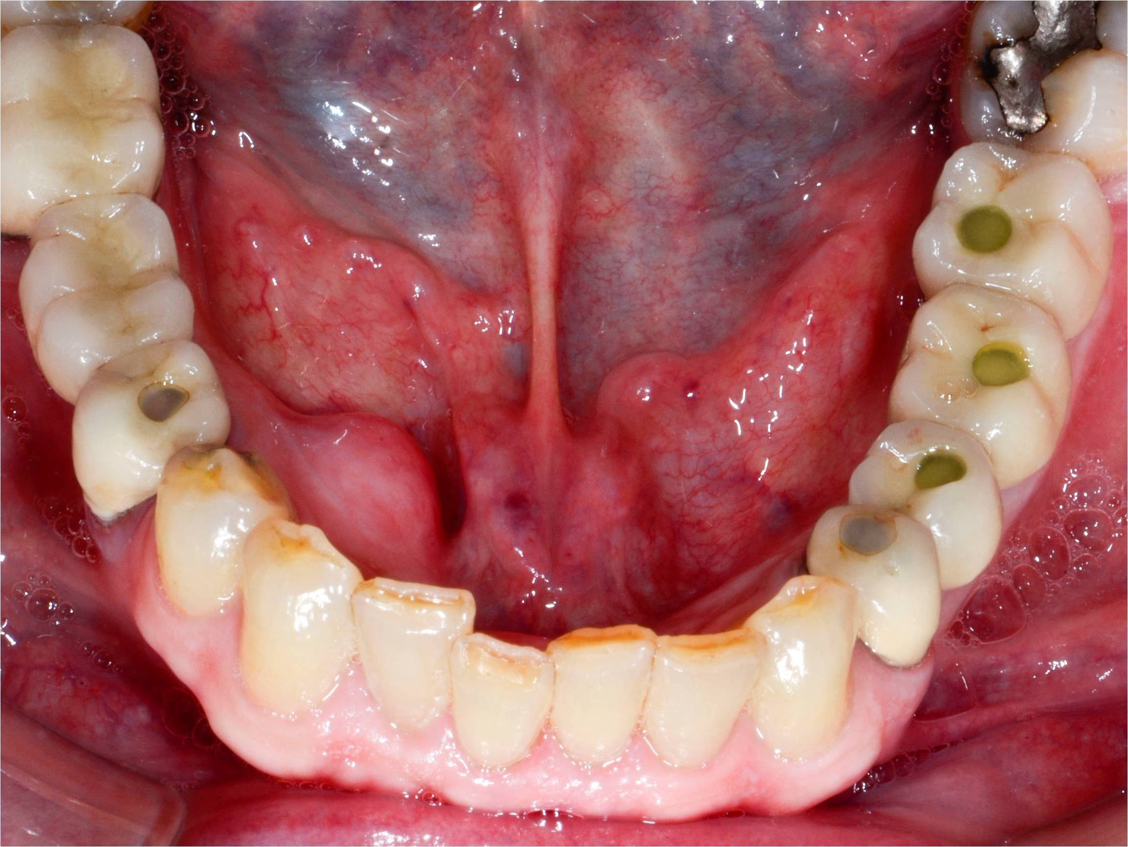
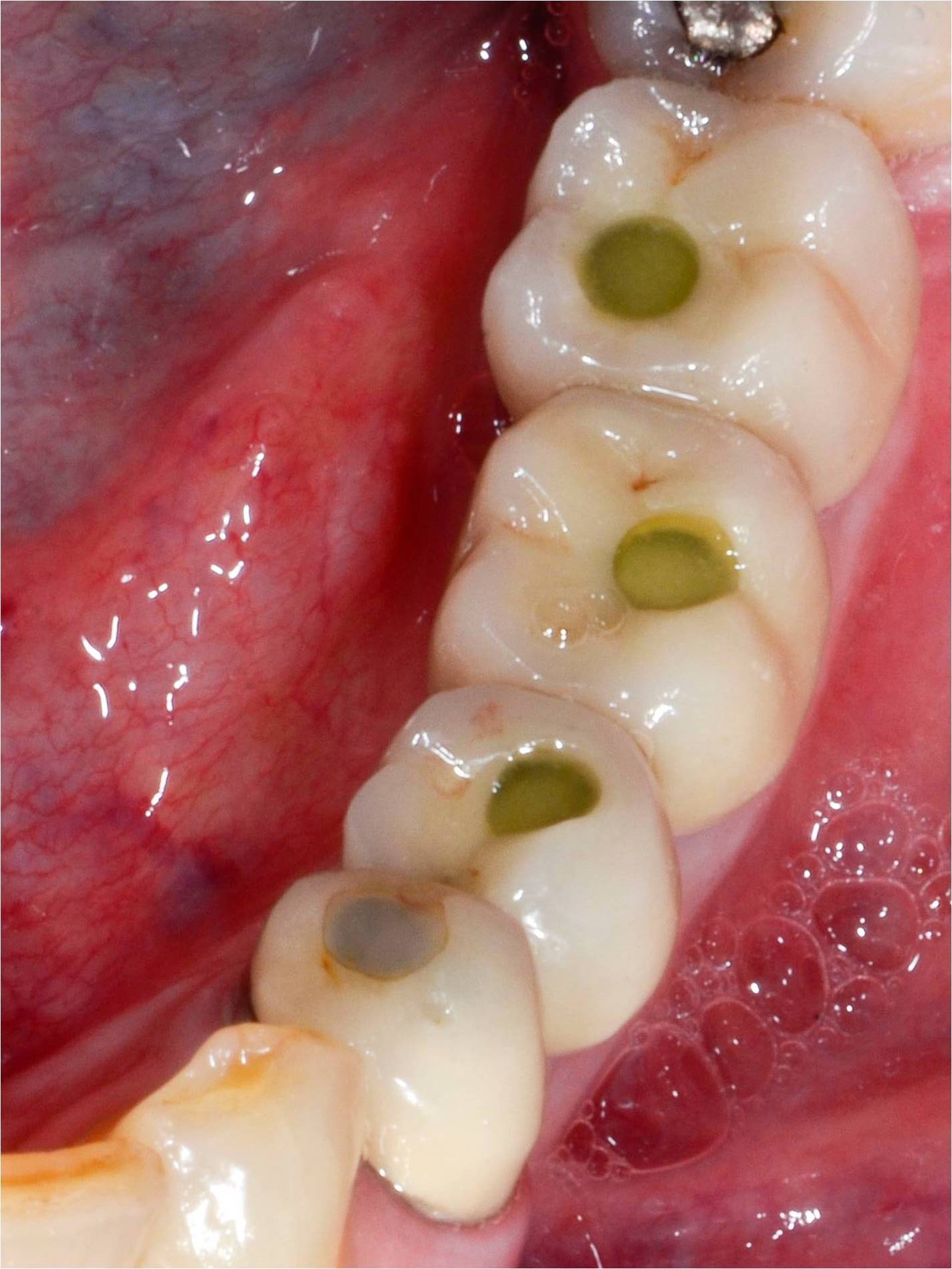
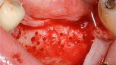
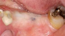
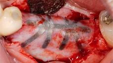
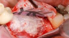
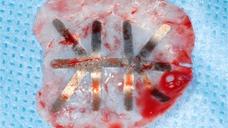
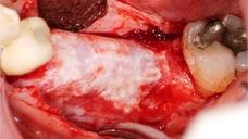
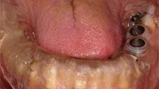
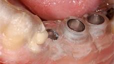
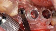
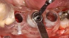
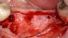
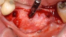
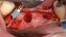
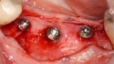
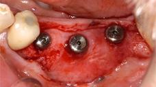
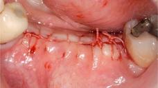
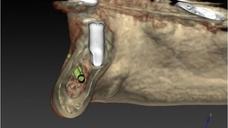
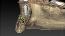
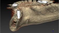
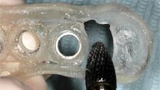
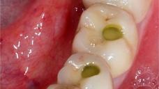
69歳男性の患者が下顎左側臼歯部に3つのヒーリングキャップが装着され、絹糸による縫合がなされた状態で来院された。2本のインプラント埋入が施術されていた。2009年上顎左側に最初にインプラント埋入を行われ、術後は疼痛がなかった。新しい歯科医による2回目のインプラント埋入後、術直後より強い痛みがあり、患者はセカンドオピニオンを求めて来院した。
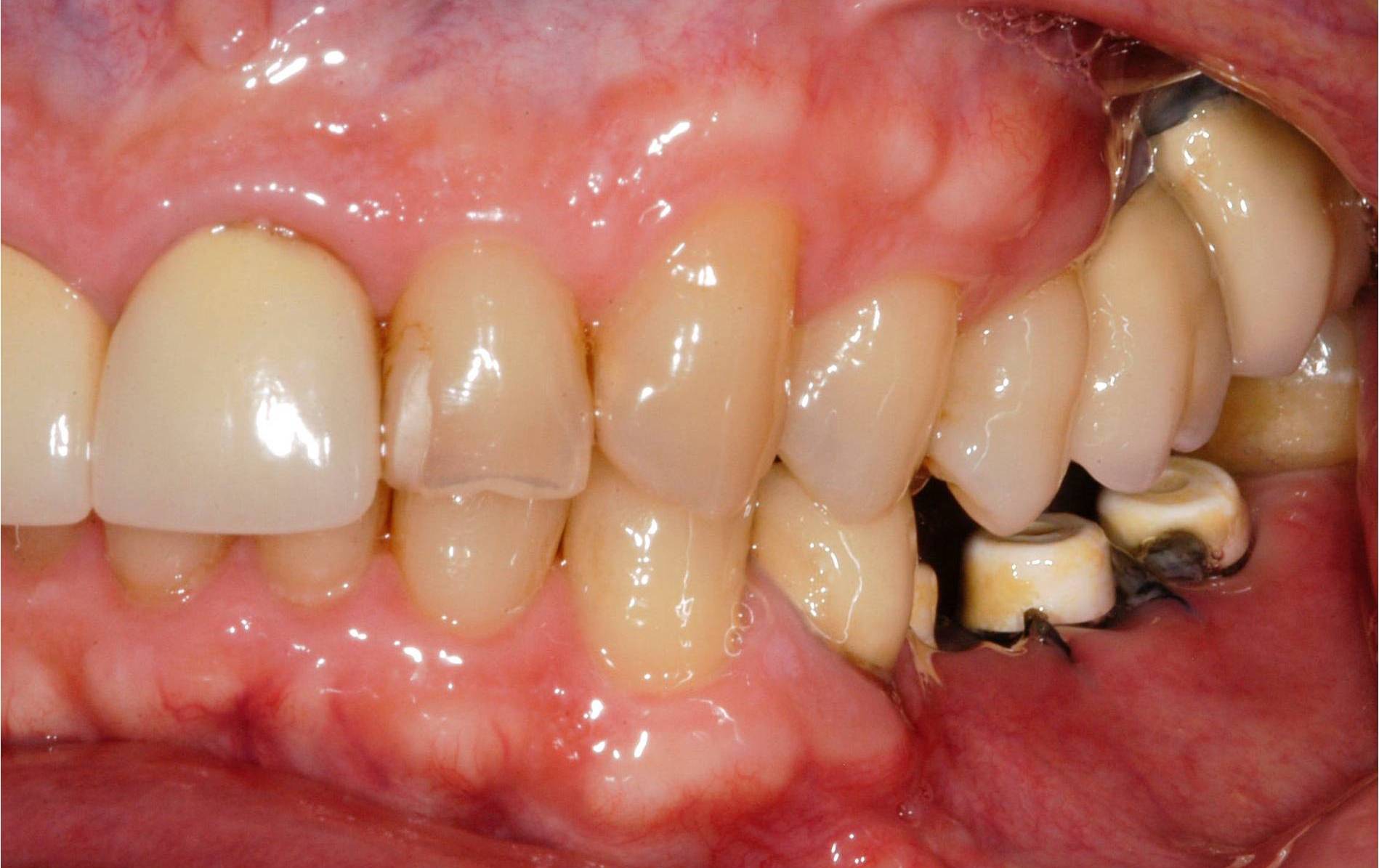
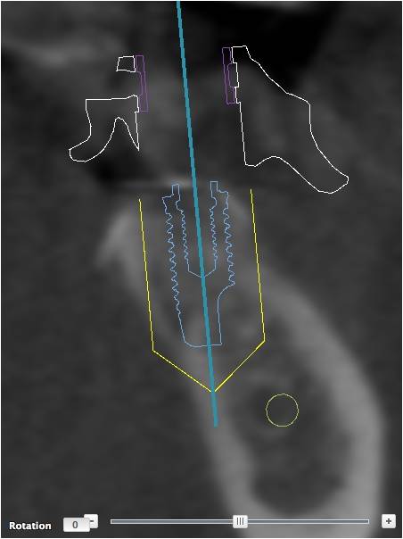
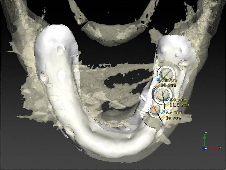
Evaluation & Diagnosis

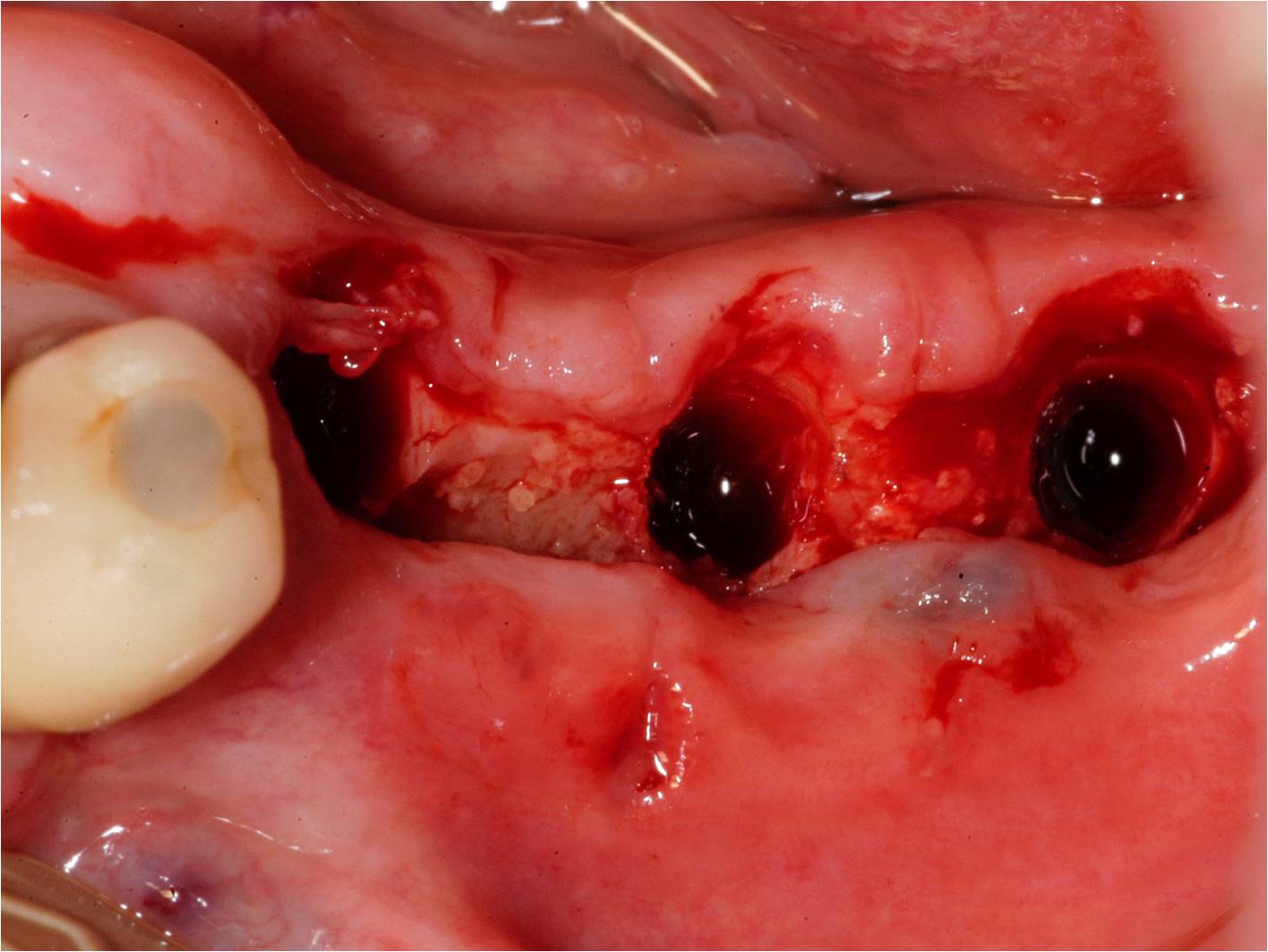
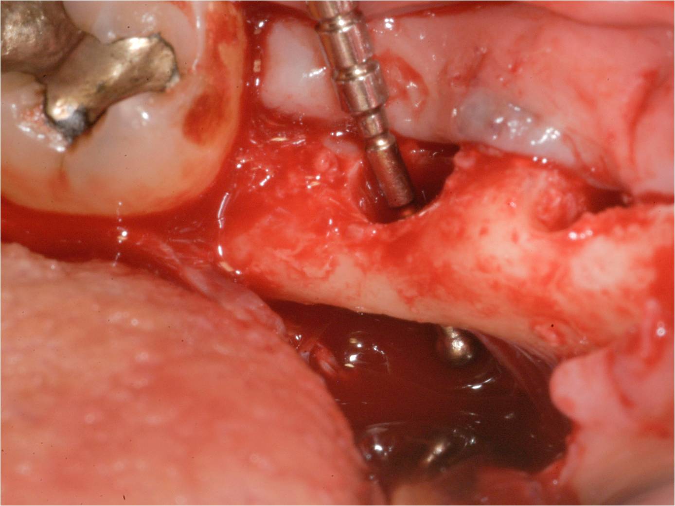
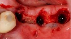
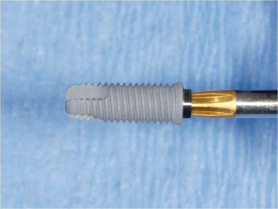
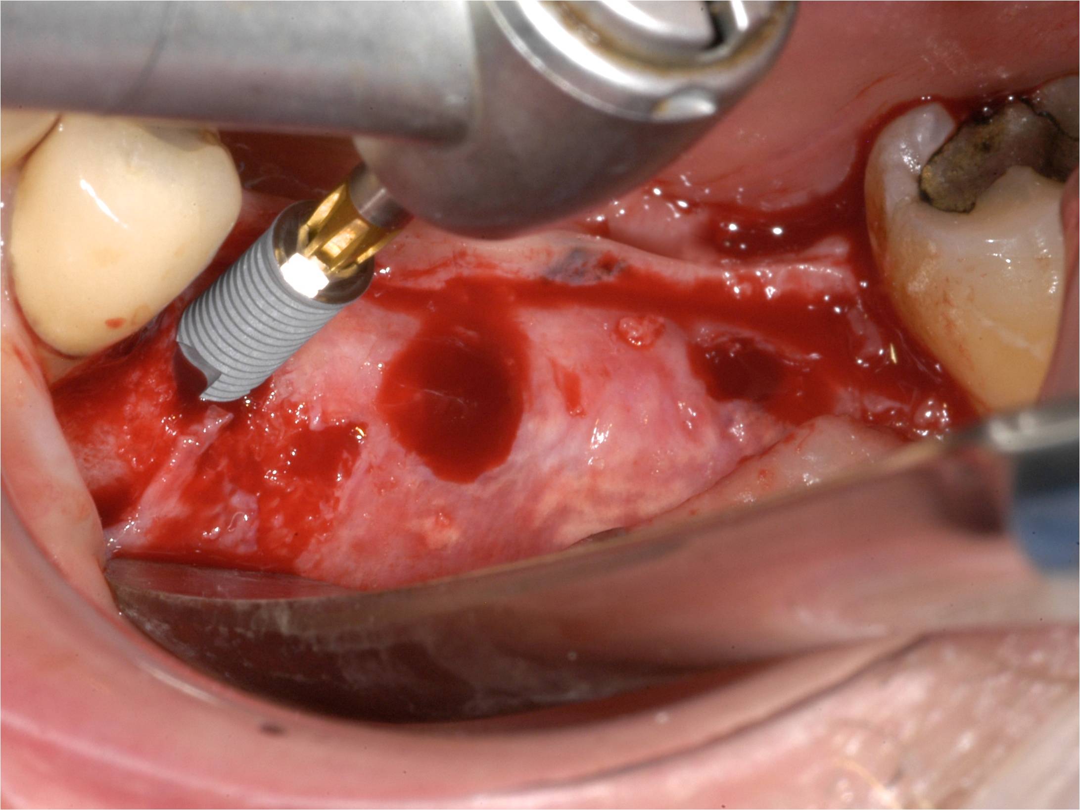
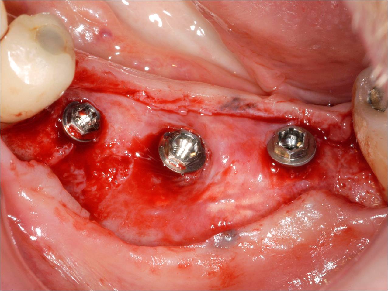
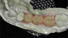
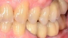
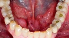
Progress & Completion


Questions
ログインまたはご登録してコメントを投稿してください。
質問する
Team members
Tom Matthes
Prosthodontist
Jerry Jacobi
Dental Technician
ログインまたは、無料でご登録して続行してください
You have reached the limit of content accessible without log in or this content requires log in. Log in or sign up now to get unlimited access to all FOR online resources.
FORウェブサイトにご登録していただきますと、すべてのオンライン・リソースに無制限にアクセスできます。FORウェブサイトへのご登録は無料となっております。